
Microscopes that are included with hardware and software systems for chromosomal analysis should meet the following basic requirements:
- adjustment of illumination using the method of Kohler;
- realized methods of contrast: light background (G,R – painting), fluorescence (Q-painting);
- three lenses with ration 10×, 40× and 100× (Oil) with the class not worse than plan-achromat;
- green light filter;
- trinocular tube with C-mount adapter with ration of 1x for installation of digital camera.
- for operation considering the method of Q-painting – a unit for realization of fluorescence method: fluorescent module; external source of light for fluorescence on the basis of metal-halide lamp, or source of light with mercury lamp, or light-emitting-diode source of light for fluorescence; filter-system for work with color DAPI.
ArgusSoft Company recommends the following models of microscopes for delivery as part of hardware-software complexes for chromosome analysis:

AxioLab A1
(Carl Zeiss). www.zeiss.com
DM 1000 & DM 1000 LED (Leica Microsystems). www.leica-microsystems.com

CX41 (Olympus).
www.olympus-lifescience.com

E200 LED (Nikon). www.nikon.com

MT 5000 (Meiji Techno). www.meijitechno.com

AxioScope A1 (Carl Zeiss). www.zeiss.com
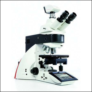
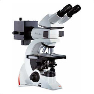
DM 2500 & DM 2500 LED (Leica Microsystems). www.leica-microsystems.com

BX43 (Olympus). www.olympus-lifescience.com

Сi-E (Nikon). www.nikon.com

MT 6000 (Meiji Techno). www.meijitechno.com
 English
 Русский